
In situ stage — within the epidermis
100% cureThe melanoma stays confined to the surface layer. A simple surgical excision is enough to cure it. This is where to act.
In 5 minutes, we photograph all of your moles in high definition. At every visit, we compare the images to detect the slightest change. Simple, painless monitoring, with no preparation needed.
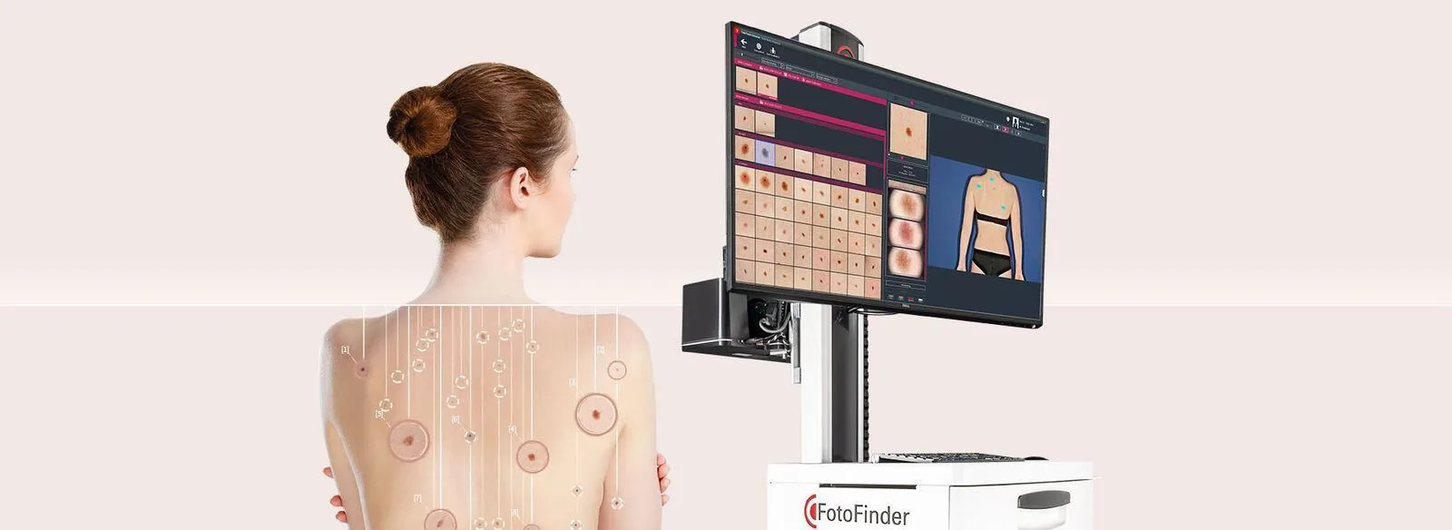
Every mole captured in high definition, then gathered and analyzed on a single screen.
of moles requiring monitoring detected automatically by our artificial intelligence.
Clinical study, EJC 2024
of cure rate when a suspicious mole is detected early.
Skin Cancer Foundation, 2024
to map all of your moles in high definition.
Fotofinder ATBM Master protocol
Fotofinder ATBM Master device, MDR-certified medical equipment used in hospital and specialized centers, in France and internationally.
CE marking · EU 2017/745






The skin is made of several layers. As long as a cancerous cell stays in the surface layer, treatment is simple and curative in more than 9 out of 10 cases. But once it crosses the basal layer and reaches the dermis, blood vessels come into play: the risk of spread begins.
That is exactly the mission of mapping: to catch every change before this critical threshold.
Simplified diagram of the skin layers. Source: standard anatomical references.
The melanoma stays confined to the surface layer. A simple surgical excision is enough to cure it. This is where to act.
Once the basal layer is crossed, the melanoma reaches the dermis and its blood and lymphatic vessels. The risk of spread begins.
Cancer cells can reach other organs (liver, lungs, brain). Treatments become far heavier and their effectiveness more uncertain.
By catching the slightest change at stage 1, Fotofinder mapping lets us act before the melanoma crosses the basal layer. That is exactly what we do for you, every year.
Dermoscan by Maison Abeille is a center dedicated to the screening and monitoring of skin cancers, built around a standard of medical quality. Here are the principles behind every exam.
Every mapping is reviewed by two physicians, in addition to the AI. Three expert perspectives to secure your diagnosis.
The analysis of your moles is performed exclusively by a physician. No nurse, no technician — never any diagnostic delegation.
Your images are hosted on a certified Health Data Hosting (HDS) infrastructure. Physician-only access, full GDPR compliance.
The team takes part in international research, attends EADV / SFD congresses, and continuously integrates advances in AI-assisted dermoscopy.
With the naked eye, tracking how skin changes stays approximate: variable lighting, imperfect memory, non-comparable manual photos. Fotofinder mapping turns this follow-up into precise, reproducible medical data that reads clearly over time.
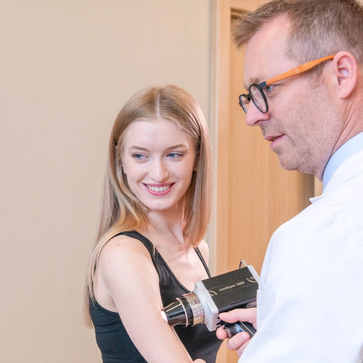
Structured monitoring dispels doubt. You leave with a clear report, a rhythm tailored to your profile, and the certainty that nothing is left to chance.
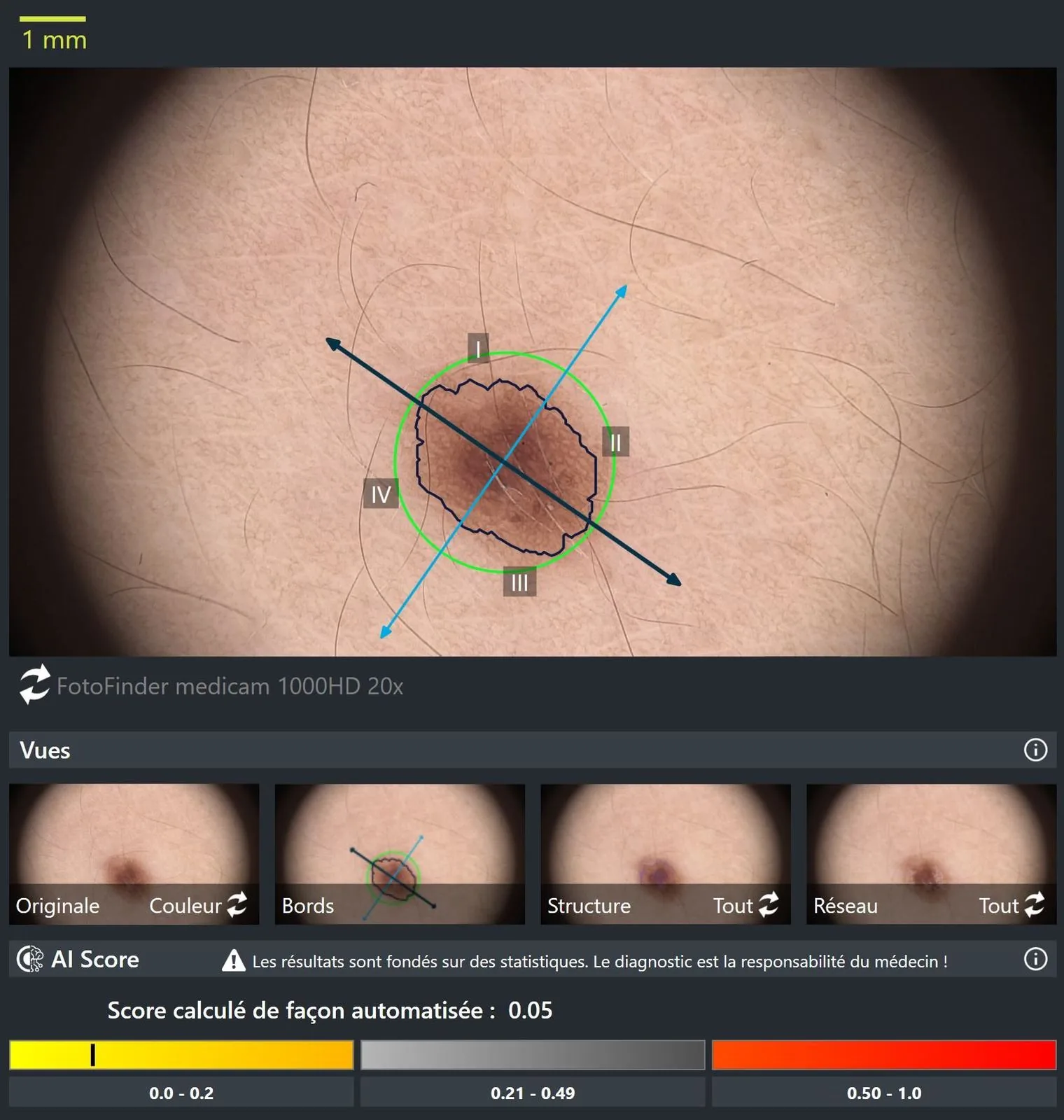
The AI examines each lesion under multiple light spectra and reveals micro-structures invisible to the naked eye. No mole is missed.
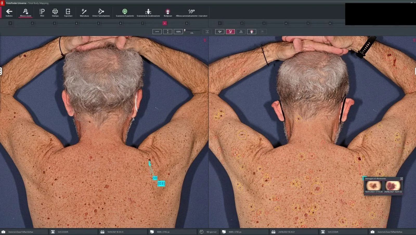
Every image is taken from the same angle, the same light, the same distance. The software overlays your visits and automatically highlights the slightest change.
This is the AI's real strength: among hundreds of moles, automatically flagging the ones that are new or changing — exactly the ones that matter.
Three ways to monitor your skin — but only one delivers the diagnostic precision of a medical device.
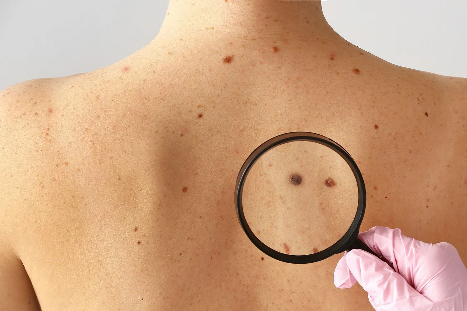
The classic dermatology check
The most common DIY follow-up
Fotofinder ATBM Master device · MDR
Fotofinder’s decisive advantage: turning approximate follow-up into reproducible, comparable, secure medical data — and enabling automatic detection of the new lesions that account for 80% of melanomas.
See exactly how a Fotofinder mapping unfolds, from check-in to report — by the Fotofinder teams who designed the device.
Official Fotofinder Systems GmbH video · for patients
No preparation required. No product applied, no pain, no radiation. You simply stand comfortably during the 5-minute mapping. The rest of the time is devoted to the dermoscopic analysis of each of your moles by your physician, plus the delivery of your report.
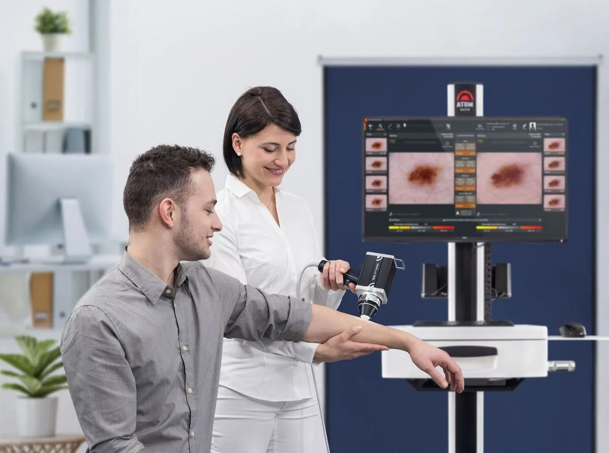
A dermatologist reviews your history and the areas you're concerned about.
The Fotofinder scanner captures all of your skin in 5 minutes, under standardized lighting.
Performed by the medical assistant
The AIMEE AI is used to detect and compare your moles over time. It performs no diagnostic evaluation: that is carried out exclusively by the physician.
You leave with a clear view of your skin’s condition and a monitoring plan tailored to your profile. No gray areas.
Every exam is performed by a physician trained in dermatology, digital dermoscopy and skin cancer screening. No diagnostic act is ever delegated to non-medical staff.
Diagnosis & dual review
Specialized dermoscopy
Reception & protocol
Our team collaborates with university research units and publishes its findings in peer-reviewed international medical journals.
Collaboration with the Center for Transplantation Sciences at Massachusetts General Hospital, affiliated with Harvard Medical School. Joint work on AI-assisted medical imaging and translational research in dermatology.
Collaboration with the Department of Plastic, Reconstructive and Aesthetic Surgery. Joint work on the role of artificial intelligence in dermatologic and plastic reconstructive surgery.
J Invest Dermatol. 2025 · doi:10.1016/j.jid.2025.12.006
Retrospective study conducted at our center, in collaboration with Cedars-Sinai Hospital (Los Angeles), Harvard Medical School / Massachusetts General Hospital (Boston), Sorbonne University, and Georges-Pompidou European Hospital (AP-HP).
Key results on 403 analyzed lesions: demonstration of the high sensitivity of dermoscopic AI in private practice, confirming the device’s ability to support early skin cancer diagnosis in real-world conditions.
lesions analyzed
USA · France · Belgique
international co-authors
Used in hospital and specialized centers, in France and internationally, the ATBM Master combines automated full-body mapping, high-definition videodermoscopy and artificial intelligence in a single EU-certified (MDR) device.
We chose it for its imaging precision and the reliability of its mole detection-and-comparison AI: AIMEE and Moleanalyzer pro. The diagnostic evaluation, for its part, is performed exclusively by the physician.
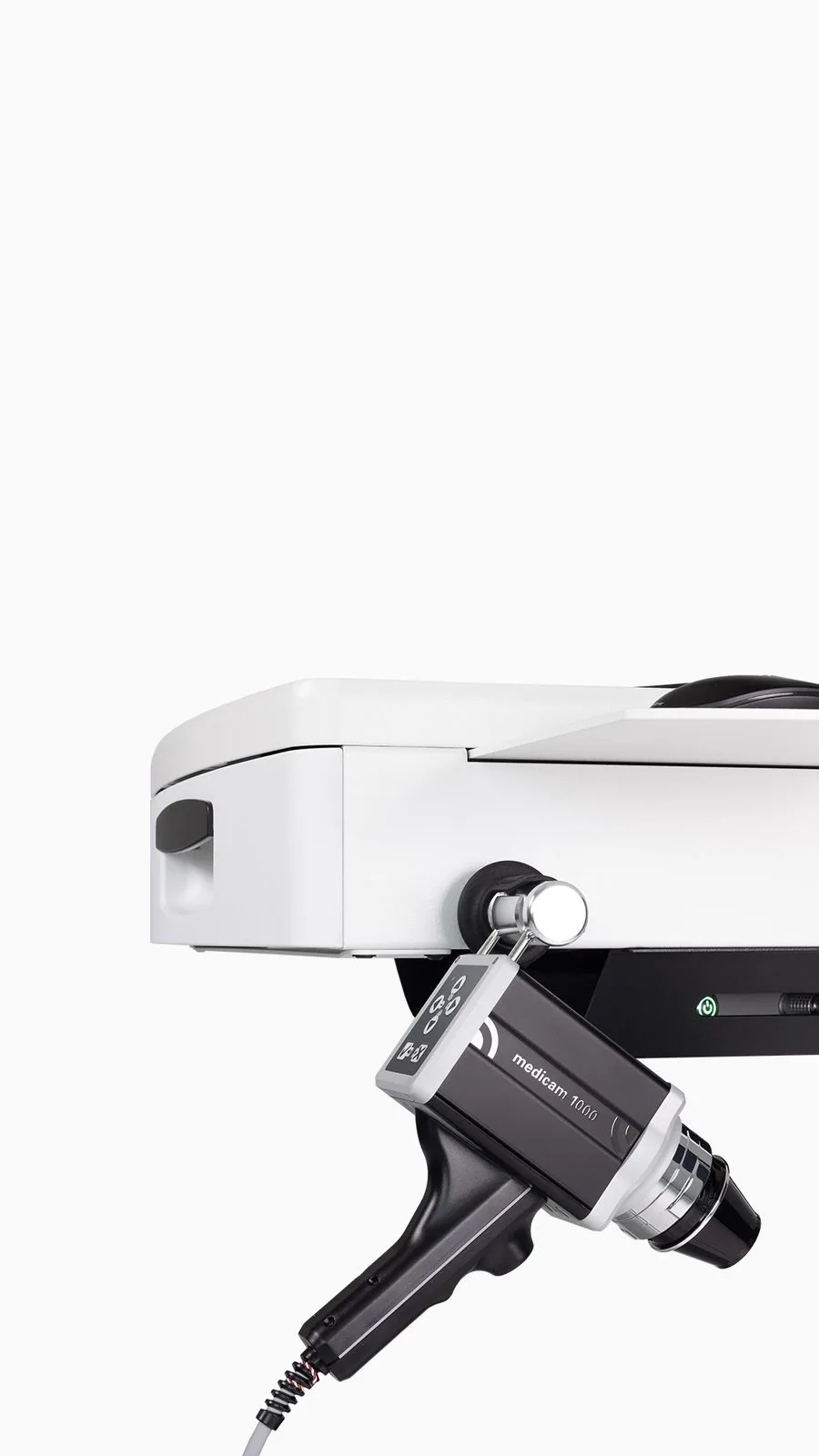
Integrated high-resolution videodermoscope to examine each lesion in detail.
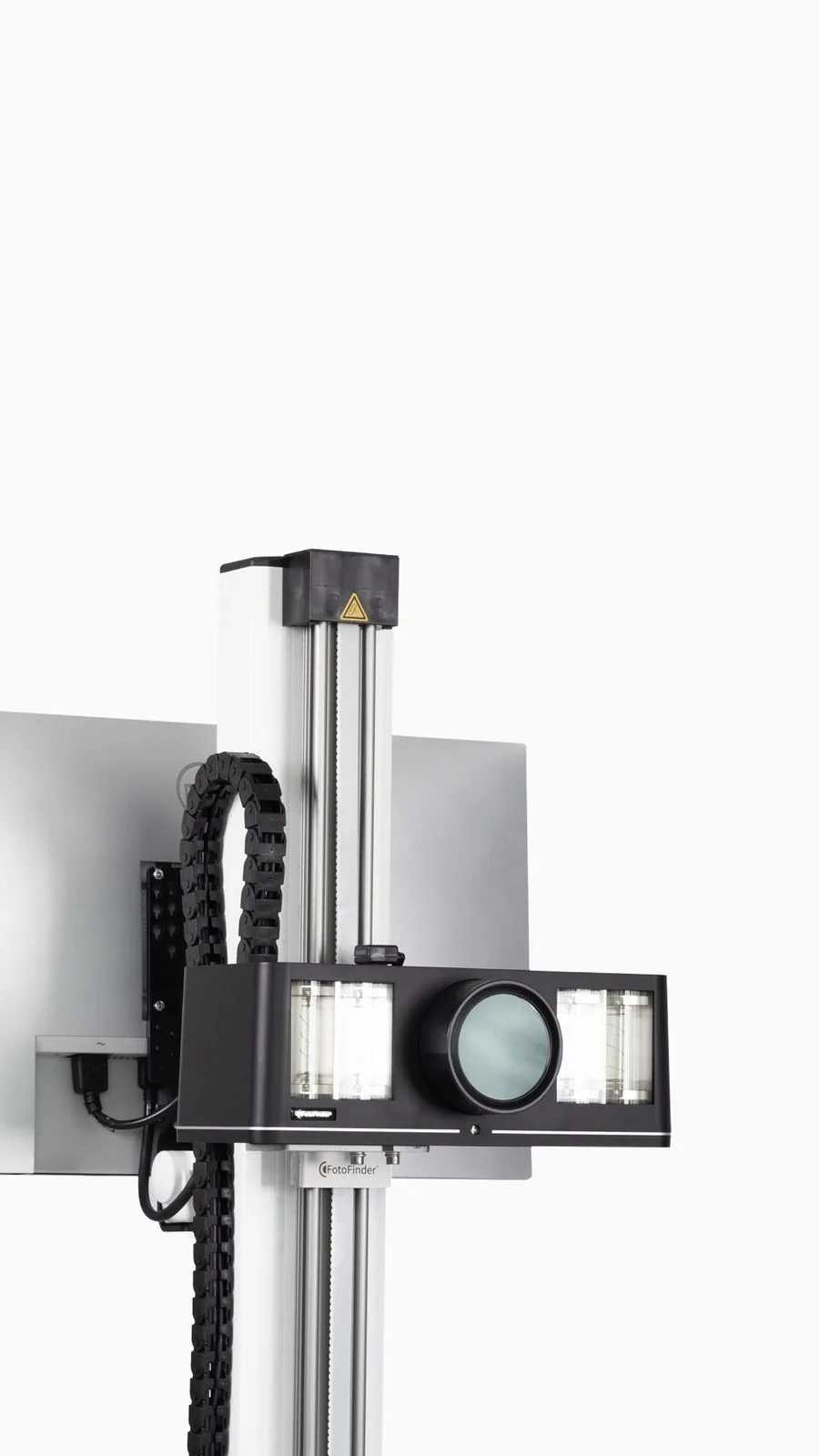
Glare-free, computer-controlled polarized light for perfectly comparable images.
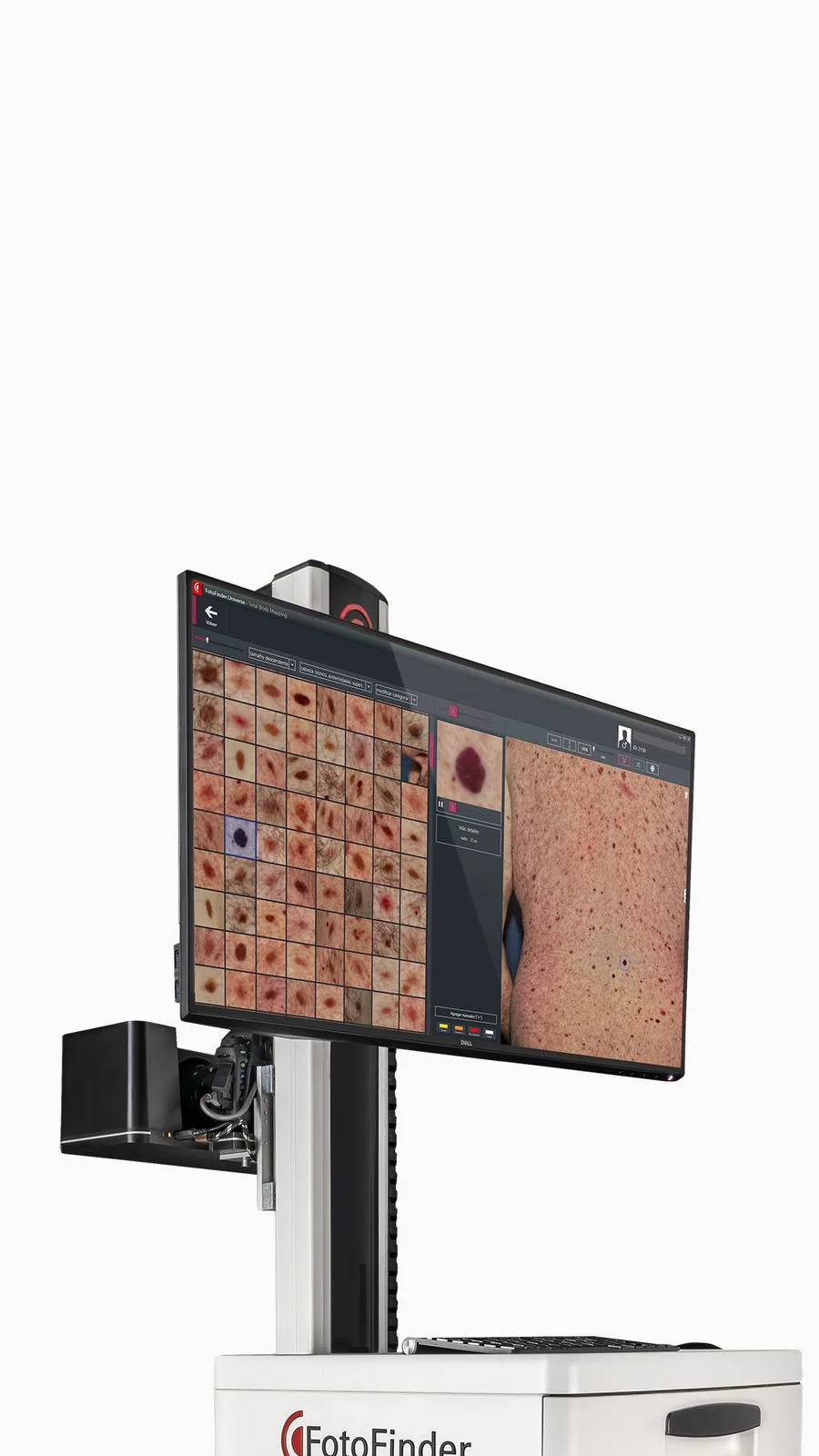
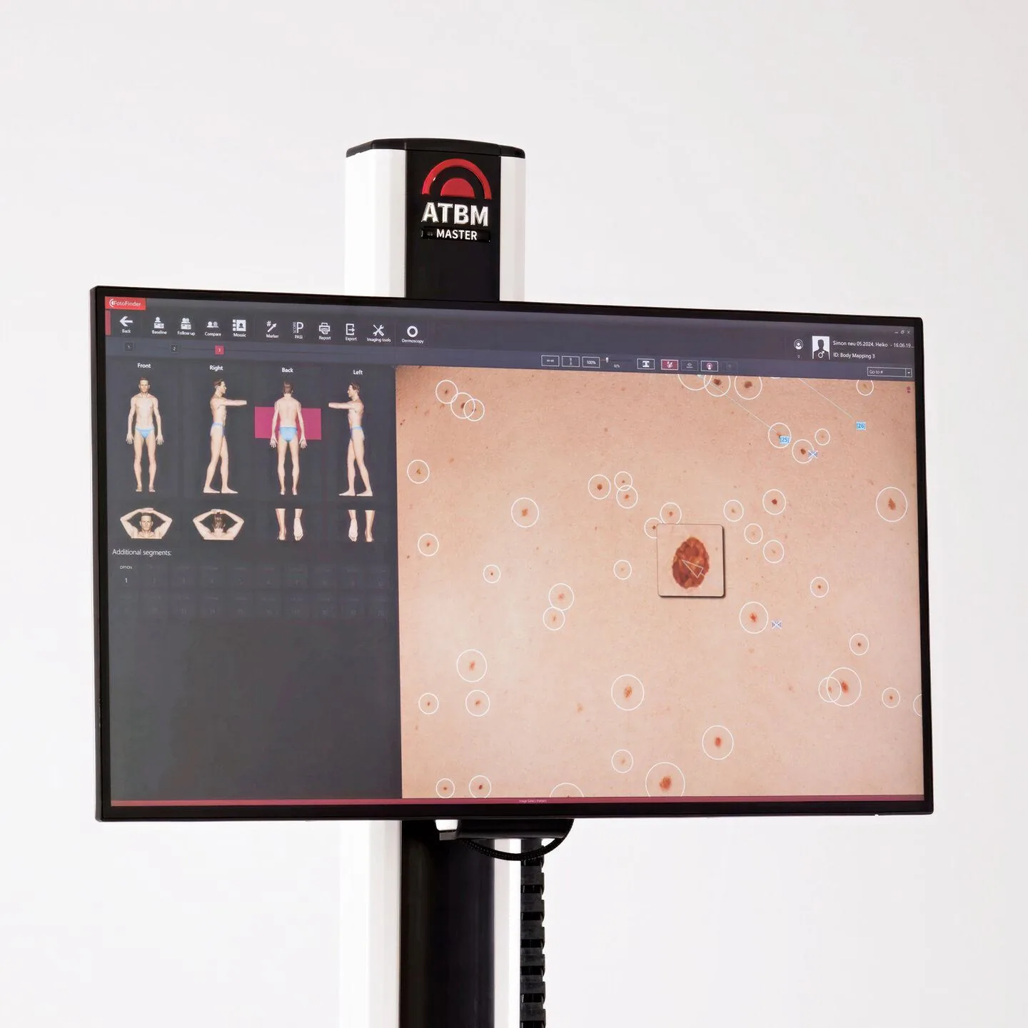
Ultra-sharp display for a precise reading of your dermoscopic images.
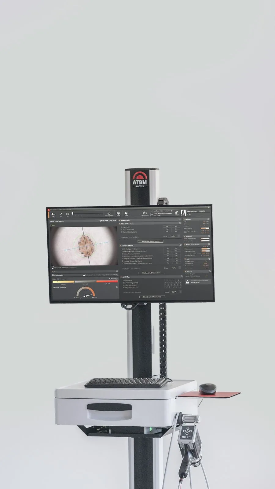
Artificial intelligence that detects and compares your moles over time. The diagnostic evaluation, for its part, remains exclusively medical.
to map your entire skin.
of lesions detected automatically during follow-up (2024 clinical study).
of unnecessary biopsies avoided thanks to AI support.
certified medical device (MDR marking).
Figures from a published clinical study (European Journal of Cancer, 2024, 403 lesions analyzed): they describe published results and do not predict an individual outcome.
Discover the ergonomics, precision and complete workflow of the Fotofinder ATBM Master device — the very same one we use at Dermoscan by Maison Abeille for every body mapping.
Official Fotofinder Systems GmbH video · available in high definition
Fully automated high-definition capture of the entire body.
PolFlash XE — glare-free medical lighting for perfectly comparable images.
Automated detection and comparison of moles over time, to assist the physician. The diagnostic evaluation remains exclusively medical.
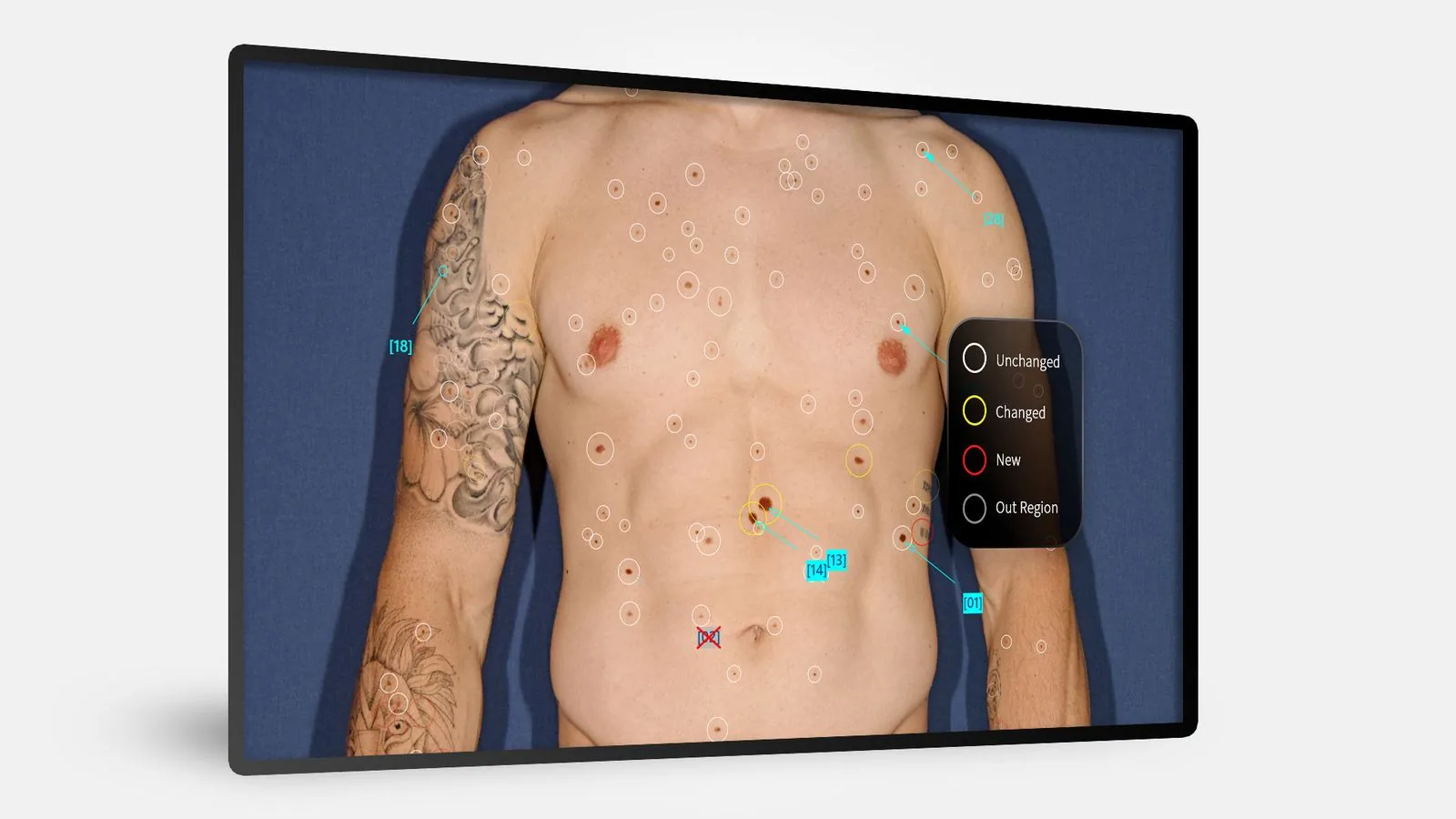
The Fotofinder Universe software automatically extracts every mole from your full-body images and arranges them in a smart mosaic, sorted into three categories: new, changed and unchanged.
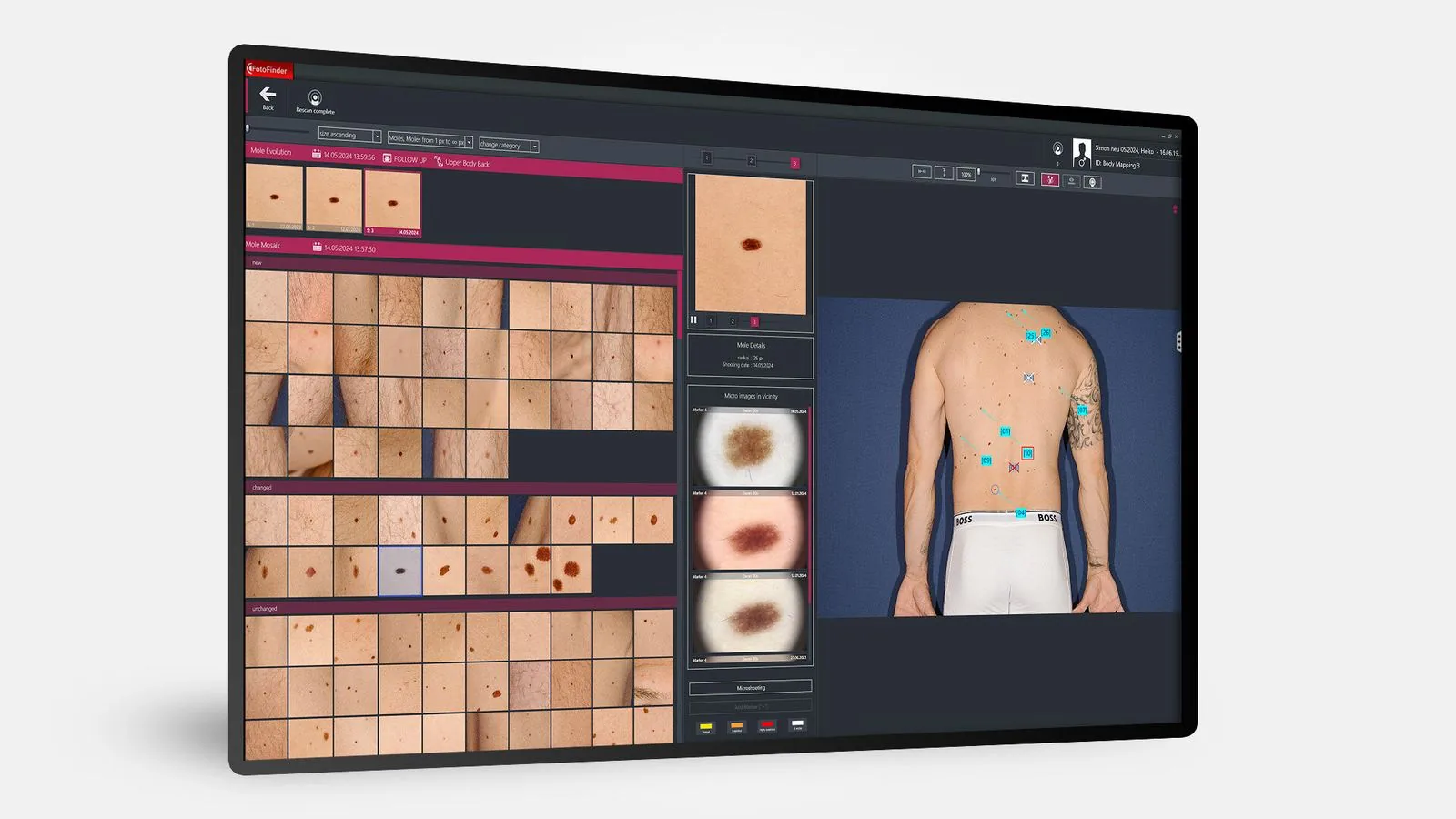
Each mole is identified and color-coded: white (unchanged), yellow (changed), red (new).
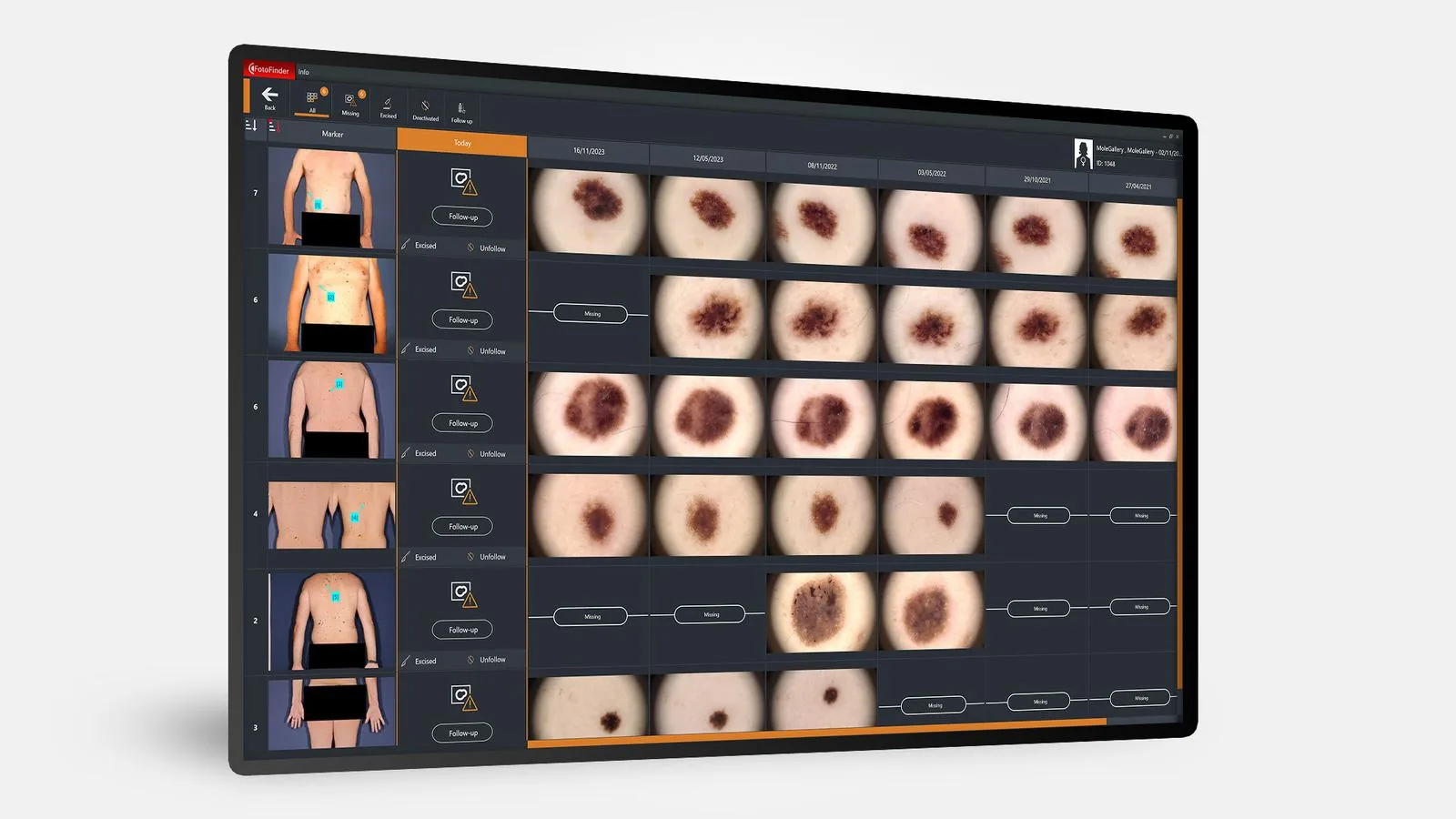
Your moles in dermoscopic close-up, in a library you can consult for life.
Our commitment is measured first by the consistency and quality of the follow-up we provide to the patients who trust us.
Two independent physicians review each mapping, in addition to the AI.
Your images are kept and compared at every visit, year after year.
The diagnostic act stays entirely in the physician’s hands. Your data and appointments are managed in-house, with no commercial intermediary.
Chiffres consolidés sur l'année 2025 · Activité du centre Dermoscan by Maison Abeille, Paris 16
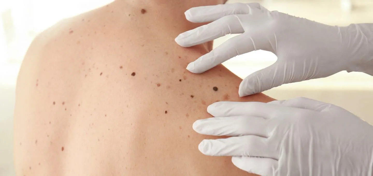
Skin mapping has no contraindications. It is especially valuable whenever you want an expert, structured eye on how your skin evolves.
A structured overview rather than a mole-by-mole check.
And anyone with frequent sun exposure.
Annual follow-up brings the peace of mind of a reliable comparison.
Anyone who wants to turn dermatology into structured monitoring.
The exam is completely non-invasive, painless and radiation-free. Suitable from adolescence and for all skin types.
A questionnaire based on recognized risk factors (Fitzpatrick phototype, number and atypia of moles, history, UV exposure). Take your time: every answer sharpens your analysis.
At the end, you get your risk level, your estimated sun capital and a personalized recommendation. Anonymous, no sign-up.
Methodology: a weighted score inspired by validated risk factors (Fitzpatrick phototype; number & atypia of nevi; personal and family history of melanoma; sunburns; artificial UV classified as a known carcinogen by IARC/WHO). Sources: WHO, IARC, American Academy of Dermatology, Skin Cancer Foundation. This self-assessment is purely indicative and is in no way a substitute for a medical exam.
Knowing your risk profile and adopting the right protective habits: two simple keys that make a real difference for your skin’s health, for life.
The risk of skin cancer rises when one or more of these factors are present. Mapping is especially valuable in these cases.
The best prevention is the kind you practice every day. Here are the golden rules — simple and effective.
Good to know: 80% of the sun damage to skin comes from cumulative exposure before age 18. Protecting your children protects their adult skin.
Clear, complete answers to 37 questions sorted by topic. Click a filter to focus on what matters to you, or browse them all.
Fotofinder full-body mapping is a medical exam that photographs all of your skin and moles to provide dermatologic monitoring that is comparative over time.
The system combines three technologies:
The images are archived to compare your skin over the years and detect any changes early.
First, the good news: when caught early, melanoma is cured in more than 9 out of 10 cases with very simple surgery. That is exactly what mapping aims for: gaining this precious time.
The earlier a lesion is detected, the more:
In France, tens of thousands of skin cancers are diagnosed every year — and the vast majority are treated effectively thanks to early detection.
The AI is a tool to support the physician, never a substitute. It helps to:
The physician makes the diagnosis. The AI adds an extra layer of precision and reliability.
A classic check relies on that day's exam, under that moment's conditions (light, angle, visual memory).
Mapping adds an irreplaceable dimension: comparison over time. It enables:
This is especially useful if you have many moles, fair skin, repeated sunburns, or a personal or family history.
Consumer apps are no substitute for a medical exam. They offer neither the imaging quality, nor the level of dermoscopic analysis, nor the medical validation of a device like Fotofinder — an EU-certified medical device (MDR) used in dermatology departments and specialized centers worldwide.
No technology guarantees 100% detection — which is exactly why mapping is a tool to support the physician, not a replacement.
Fotofinder considerably improves the quality of follow-up and detection. A definitive diagnosis may sometimes require close monitoring, a biopsy, or a surgical excision with laboratory analysis.
Yes. Contrary to a common belief, melanoma risk is not limited to skin with very many moles.
About 70 to 80% of melanomas appear on a new lesion, not on an existing mole. Mapping therefore creates a 'reference photo' of your skin at a given moment. At later checks, the system instantly spots any new or changed lesion, even a very subtle one.
Even with few moles, this comparative follow-up can make all the difference.
Yes — it's one of the recognized factors. Having more than 50 moles, especially atypical ones, significantly raises the risk compared with the general population.
In that case, mapping becomes especially valuable: tracking 50 moles with the naked eye is nearly impossible, whereas the system does it effortlessly.
Yes. People with red or fair hair, light eyes, freckles, or who burn easily in the sun statistically have a higher risk of skin cancers.
In red-haired people carrying certain genetic variants (the MC1R gene), the risk can be even more pronounced. Mapping then offers real peace of mind.
Very often, yes. A family history — especially among first-degree relatives (parents, siblings) — raises the risk of developing a skin cancer. The structured, comparative follow-up of mapping is a particularly well-suited prevention tool in this case.
Yes — it's even a major indication. After a first melanoma or skin cancer, the risk of developing a second is higher than in the general population. Fotofinder's comparative digital follow-up is especially relevant here — it's the reference tool for this type of monitoring.
Yes. Melanoma can progress for a long time without pain or obvious signs — which is exactly why regular follow-up makes it possible to catch it at a very early stage, when treatment is simple and quick.
It depends on the medical context. Mapping is generally offered to adults, but it can be relevant for a child or teenager with a significant family history, atypical nevi, or an at-risk background. We tailor the indication to each situation.
Absolutely. It's actually a very healthy approach. Many patients consult without any serious lesion, simply to get a precise baseline, secure their follow-up, and have an 'ID card' of their skin for the years ahead. That's exactly what mapping is for.
Yes. Skin cancers can also affect darker phototypes, even if some forms are less frequent. Presentations may differ depending on phototype — hence the importance of specialized medical expertise and quality mapping, like the one we provide.
Yes. Cumulative UV exposure — and above all severe sunburns in childhood and adolescence — is one of the major risk factors for skin cancer. A few significant sunburns are enough to meaningfully raise future risk, even decades later.
On average 30 to 60 minutes depending on the number of lesions and the type of follow-up, including only 5 minutes of automated body scan. The rest of the time is devoted to the consultation, targeted dermoscopic analysis, and the delivery of a report.
Not at all. The exam is completely painless: no injection, no product applied, no harsh contact. It involves only medical photography and dermoscopic analysis.
None. Fotofinder uses:
It is simply a medical photographic imaging system using visible light.
Avoid heavy makeup on the areas to be analyzed, as well as very greasy creams applied just before the session. Dark nail polish can sometimes hinder nail examination if nail monitoring is needed. Otherwise, no special preparation.
Mapping requires good visualization of the skin to be reliable. The physician adapts the exam with respect, discretion, and to your comfort. You keep your underwear on, and every step is explained in advance.
Only if necessary, or if you yourself report a lesion in that area. Some lesions can appear in less visible areas, which is why the option exists — but it remains at your initiative and with your explicit consent.
Yes — and that is precisely the point of comparative follow-up. Your medical images are securely archived in your patient record, in accordance with French health-data regulations, to allow reliable comparison at your future checks.
Only the authorized healthcare professionals involved in your care, in strict compliance with medical confidentiality and French health-data regulations (GDPR, HDS hosting).
Fotofinder full-body mapping is generally considered an out-of-schedule (hors nomenclature) procedure. It is therefore usually not reimbursed by the French national health insurance. Some private health plans (mutuelles) do offer partial coverage — feel free to check with yours.
The fee reflects the level of rigor and medical quality we commit to on every exam. Four elements make up this cost:
Every mapping is analyzed by two physicians: your referring dermatologist and a second physician for the dual review. This deliberate redundancy is what truly secures the diagnosis — it's the standard applied in the best screening centers.
The dermoscopic analysis of your moles is performed exclusively by a physician. No nurse, no assistant, no technician or non-medical staff ever places a diagnostic eye on your images. It's non-negotiable, and it's what sets us apart from centers where dermoscopy is delegated.
During your visit, you are supported by a trained team: medical assistants dedicated to the image-capture protocol, a medical secretariat to manage your record, and physicians for the exam and the dual review. Everything is organized so your visit is smooth and your time is respected.
Fotofinder ATBM Master (one of the most advanced medical devices in the world), secure HDS archiving of your images for lifelong follow-up, clinically validated AI for mole detection and comparison, and significant physician time devoted to the consultation and report.
The fee is communicated to you transparently when you book. Some private health plans offer partial coverage.
Yes, with no contraindication whatsoever. Mapping is:
Pregnancy can in fact change some moles — so monitoring can be especially useful during this period.
Yes, completely. Mapping is compatible with breastfeeding, with no constraints at all.
Virtually none. Mapping is compatible with:
Since there is no radiation, no injection, and no invasive act, the exam is considered very safe.
It's not ideal. An intense tan or inflamed skin (redness, peeling) can make analysis more delicate. If possible, wait a few weeks after heavy exposure for your skin to return to its usual state — your images will be more usable.
No, don't wait. Any rapid change in:
warrants medical advice without delay. Contact us: we'll find a suitable slot to examine that lesion as a priority.
Not always, and that's normal. Some lesions require:
Melanoma medicine often relies on analyzing change over time — and that's exactly what mapping does with unmatched precision.
Depending on the situation, several options are possible:
The goal is twofold: avoid diagnostic delays, but also avoid unnecessary surgeries. Fotofinder's precision is precisely what helps reduce superfluous biopsies.
Yes. If a lesion looks concerning, surgical management can be arranged quickly, either at the center or together with the surgeons we usually work with. We support you at every step.
No, that's a stubborn myth. A melanoma can be:
This is one reason dermoscopic analysis and Fotofinder's AI are so valuable: they catch what a visual exam might miss.
It depends on your risk level and is determined medically after your first exam. Depending on the case:
You always leave with a clear, personalized monitoring plan.
Absolutely. Your images are kept in your secure medical record, accessible only to the medical team caring for you, in compliance with GDPR and French regulations on health-data hosting (HDS certification).
Still have a question?
Contact us directlyThe Dermoscan by Maison Abeille center is located on Avenue du Colonel Bonnet, a few steps from Trocadéro. A calm, bright space entirely dedicated to advanced dermatology.
To offer you a truly tailored exam, we need to get to know you a little. 9 quick questions, then you go straight to our available slots.
Book your skin mapping online : choose your slot directly in our secure calendar.
Choose a slotA medical qualification discussion takes place before or during the consultation. No health data is collected through this website : your medical information is gathered in a secure setting, at the center.